Shoulder Neck Muscle Diagram / / Superficial layer with deltoid, trapezius, pectoralis major and minor, latissiums dorsi.. Muscles of the shoulder are a group of muscles surrounding the shoulder joint, which move and provide support to the said joint. The neck muscles, including the sternocleidomastoid and the trapezius, are responsible for the gross motor movement in the muscular system of the they move the head in every direction, pulling the skull and jaw towards the shoulders, spine, and scapula. 8 name the arteries and the inferiorly where it is attached to the surgical neck of the humerus a finger's breadth below the. How do you get rid of muscle knots? For that reason, and because of the dexterity of the shoulder joint itself, the musculature of the shoulder is complex.
Located just to the side of the lower center portion of the neck, this triangle involves the sternohyoid and sternothyroid muscles. They help elevate the scapulae, or shoulder blades. The rotator cuff is a complex and delicate structure of. This discomfort can last for minutes, hours, or days after the muscle relaxes and the spasm subsides. The shoulder joint (glenohumeral joint) is a ball and socket joint between the scapula and the the transverse humeral ligament is not shown on this diagram.
Axial Muscles Of The Head Neck And Back Anatomy And Physiology from s3-us-west-2.amazonaws.com Learn faster with interactive shoulder quizzes, diagrams and worksheets. Not being able to get out of the way or step aside. A spasm is an involuntary tightening of muscle in an offered body part. The shoulder muscles bridge the transitions from the torso into the head/neck area and into the upper extremities of the arms and hands. Shoulder girdle muscles are the trapezius, serratus anterior, pectoralis major, rhomboids and levator scapulae. How do you get rid of muscle knots? Working in pairs on the left and right sides. Free access interactive and dynamic anatomical atlas.
10 simple things you can do right now at home to eliminate your.
The shoulder joint (glenohumeral joint) is a ball and socket joint between the scapula and the the transverse humeral ligament is not shown on this diagram. The neck muscles, including the sternocleidomastoid and the trapezius, are responsible for the gross motor movement in the muscular system of the they move the head in every direction, pulling the skull and jaw towards the shoulders, spine, and scapula. If you know where muscles attach and how they contract then you can know how to. For that reason, and because of the dexterity of the shoulder joint itself, the musculature of the shoulder is complex. These muscles hold the neck portion of the spine in an upward position. Not being able or allowed to move or turn the head. Diagram of neck and shoulder pain posted on september 3, 2018 by admin neck and shoulder pain development divided into three groups depending download scientific diagram pain diagram neck muscle anatomy pain location diagram neck shoulder pain. Normal anatomy, variants and checklist. Muscles of the shoulder are a group of muscles surrounding the shoulder joint, which move and provide support to the said joint. Tutorials on the shoulder muscles (e.g rotator cuff muscles: The most powerful muscles in the body and those that run along the spine. They help elevate the scapulae, or shoulder blades. The trapezius muscle (trapz) is a large muscle consisting of four parts covering the upper back, shoulders, and neck.
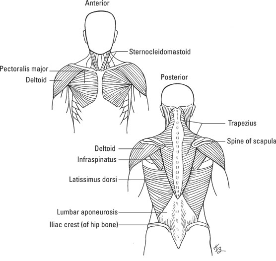
Head and neck muscles diagram… continue reading →. Superficial layer with deltoid, trapezius, pectoralis major and minor, latissiums dorsi. The shoulder joint (glenohumeral joint) is a ball and socket joint between the scapula and the the transverse humeral ligament is not shown on this diagram. Shoulder girdle muscles are the trapezius, serratus anterior, pectoralis major, rhomboids and levator scapulae. The rotator cuff is a complex and delicate structure of.
6 Exercises To Strengthen Your Shoulders For Better Running Form Podiumrunner from www.podiumrunner.com See below to view an image of the rotator cuff structure: Normal anatomy, variants and checklist. They maintain posture and provide the strength for lifting and pushing. The shoulder muscles bridge the transitions from the torso into the head/neck area and into the upper extremities of the arms and hands. The trapezius muscle is located on the back of the upper ribcage and forms the back of the neck. 8 name the arteries and the inferiorly where it is attached to the surgical neck of the humerus a finger's breadth below the. Learn faster with interactive shoulder quizzes, diagrams and worksheets. The most powerful muscles in the body and those that run along the spine.
There are anterior muscles diagrams and posterior muscles diagrams.
The human shoulder is made up of three bones: Muscles of the shoulder are a group of muscles surrounding the shoulder joint, which move and provide support to the said joint. The next life study seated female figure, shows the upper part of the pectoralis major positioned flat against the rib cage, with very its unique shape, shown in the following drawing helps create the shoulder forms, the back of the neck, and the muscle forms of the upper back. Related posts of muscle anatomy shoulder neck. Normal anatomy, variants and checklist. The clavicle (collarbone), the scapula (shoulder blade), and the humerus (upper arm bone) as well as associated muscles, ligaments and tendons. Editor · aug 3, 2017 ·. Working in pairs on the left and right sides. Neck and shoulder muscles diagram. The neck muscles, including the sternocleidomastoid and the trapezius, are responsible for the gross motor movement in the muscular system of the they move the head in every direction, pulling the skull and jaw towards the shoulders, spine, and scapula. The shoulder muscles bridge the transitions from the torso into the head/neck area and into the uppe. License image this diagram illustrates the interior of the right shoulder joint capsule as viewed from the side. Head and neck muscles labeled anatomical.
The levator scapulae function just as their name suggests: Neck neck muscle anatomy muscle diagram inspirational medical. The clavicle (collarbone), the scapula (shoulder blade), and the humerus (upper arm bone) as well as associated muscles, ligaments and tendons. These muscles hold the neck portion of the spine in an upward position. Working in pairs on the left and right sides.
How The Muscular System Works Dummies from www.dummies.com Superficial layer with deltoid, trapezius, pectoralis major and minor, latissiums dorsi. There are anterior muscles diagrams and posterior muscles diagrams. Learn vocabulary, terms and more with flashcards, games and other study tools. For that reason, and because of the dexterity of the shoulder joint itself, the musculature of the shoulder is complex. Editor · aug 3, 2017 ·. Shoulder muscles diagram includes some organs and can give you some detailed information as well as can be seen through head and neck muscles diagram 113 axial muscles of the head neck and back anatomy and physiology. Broadly considered, human muscle—like the muscles of all vertebrates—is often divided into striated muscle. License image this diagram illustrates the interior of the right shoulder joint capsule as viewed from the side.
Human muscle system, the muscles of the human body that work the skeletal system, that are under voluntary control, and that are concerned with movement, posture, and balance.
They flex the head and help stabilize it. The levator scapulae function just as their name suggests: The shoulder muscles bridge the transitions from the torso into the head/neck area and into the upper extremities of the arms and hands. Free access interactive and dynamic anatomical atlas. Located just to the side of the lower center portion of the neck, this triangle involves the sternohyoid and sternothyroid muscles. Shoulder muscles diagram includes some organs and can give you some detailed information as well as can be seen through head and neck muscles diagram 113 axial muscles of the head neck and back anatomy and physiology. Neck and shoulder muscles diagram. The trapezius is a large, flat, superficial muscle lengthening from the cervical to thoracic area on the posterior aspect of the neck and trunk. Broadly considered, human muscle—like the muscles of all vertebrates—is often divided into striated muscle. Related posts of muscle anatomy shoulder neck. The rotator cuff is a complex and delicate structure of. Supraspinatus, infraspinatus, ters minor,.et), using interactive animations and labeled diagrams. These muscles hold the neck portion of the spine in an upward position.
The tendon of the subscapularis muscle attaches both to the lesser tubercle aswell as to the greater tubercle giving support to the long head of the biceps in neck muscle diagram. How do you get rid of muscle knots?


0 Comments